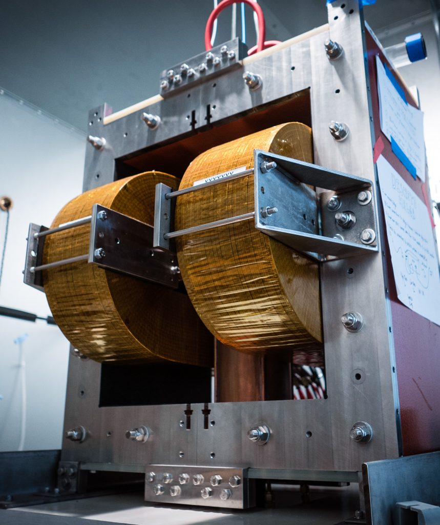
Magnetic Particle Imaging (MPI) is an emerging tracer medical imaging technology. Unlike an x-ray, MRI or CT scan, tracer technologies such as PET, SPECT and MPI do not image tissues and bones. Instead, they track the location of an injected tracer throughout the body, enabling high contrast imaging of bodily functions. Tracer technologies are fundamental in hospitals today to assess heart function, diagnose cancer, find sources of inflammation, among many other applications. MPI offers an ionizing radiation free alternative, with potential for new contrast mechanisms via nanoparticle relaxation and high-resolution imaging via new imaging pulse sequences and hardware.
How does it work? MPI uses superparamagnetic iron oxide nanoparticles (SPIOs) as tracers. To run an MPI scan, an excitation field is applied to SPIOs in the field-of-view (FOV). The magnetic dipoles reorient rapidly in response to the excitation field. Much like in MRI, the change in magnetization can be visualized via Faraday’s law with a receiver coil. Unlike MRI, the change is of electronic magnetization, rather than nuclear magnetization. This results in high MPI sensitivity to SPIOs because the electronic magnetization of iron is 22 million times stronger than that of the nuclear magnetization of water at 7 Tesla. To localize this signal, a large gradient field is used. Outside of a small region with a close to zero field, termed the field free region (FFR), the gradient locks SPIOs in place even if the excitation field is applied. Inside the FFR, the SPIOs reorient in response to the excitation field. By rastering this FFR across each point in the FOV, an image is created.